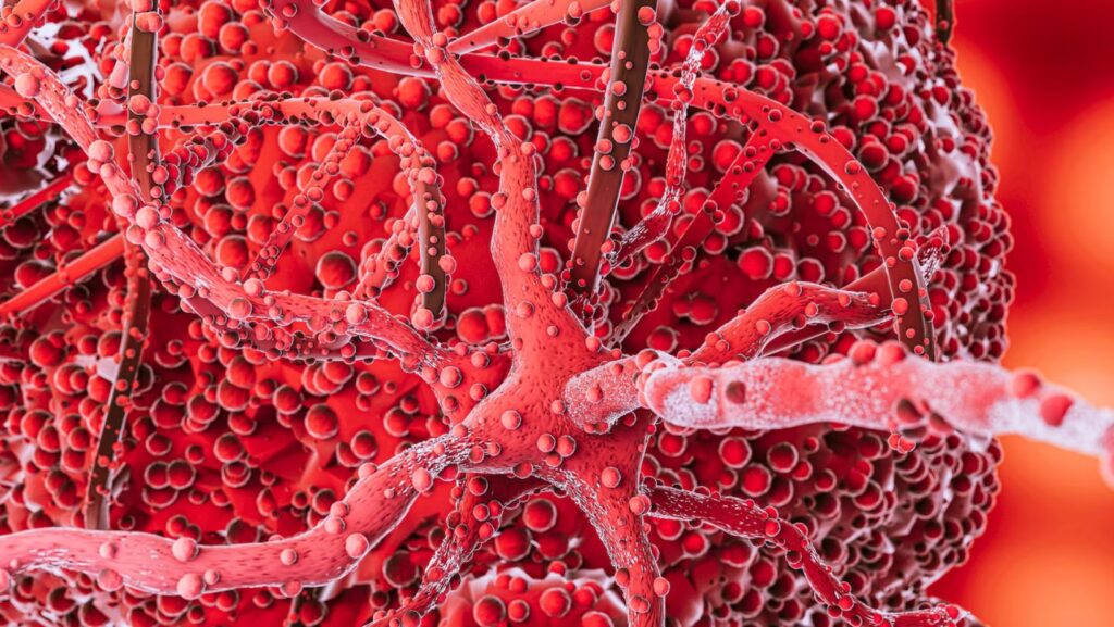
A groundbreaking study from the UConn School of Medicine has revealed a critical cellular malfunction that could fundamentally shift how scientists understand — and eventually treat — devastating brain disorders like Alzheimer’s disease, amyotrophic lateral sclerosis (ALS), and frontotemporal dementia (FTD). The study, published March 14 in Nature Neuroscience, focuses on the brain’s blood vessels and their key role in driving Neurodegenerative Diseases.
Blood Vessels Take Center Stage: Neurodegenerative Diseases

For decades, neurodegenerative research has mainly focused on neurons — the brain’s information-processing cells — while the brain’s blood vessels were often viewed as little more than passive plumbing. But the new findings challenge that narrative.
Led by MD/Ph.D. candidate Omar Moustafa Fathy, who works under vascular biologist Dr. Patrick A. Murphy, the research shines a spotlight on the brain’s vascular system—specifically, the endothelial cells that line blood vessels and form the central component of the blood-brain barrier (BBB). The BBB plays a vital defensive role, shielding the brain from harmful substances circulating in the bloodstream. However, when that barrier breaks down, the brain becomes vulnerable to inflammation and degeneration.
“It’s often said we’re only as old as our arteries,” Murphy noted. “Across diseases, we are learning the importance of the endothelium. I had no doubt the same would be true in neurodegeneration.”
A Closer Look at Elusive Cells
One major hurdle in this line of research is that endothelial cells are rare and notoriously difficult to isolate, especially from the frozen tissue samples typically stored in biobanks. To solve this, Fathy and the team pioneered a new method to enrich these cells from frozen human brain tissue. They then applied a powerful tool called inCITE-seq, marking the first time it has ever been used in human samples, to measure protein-level signaling in single cells.
This approach allowed researchers to analyze blood vessel cells from patients with Alzheimer’s, ALS, and FTD and compare them directly with samples from healthy aging individuals.
The result? Across all three diseases, endothelial cells showed a shared and previously unidentified signature of dysfunction — including a consistent loss of TDP-43, a protein already known to play a role in ALS and FTD and increasingly recognized in Alzheimer’s pathology. Until now, TDP-43 depletion had been seen mainly in neurons, not the vascular system.
A New Direction for Diagnosis and Treatment
The study’s findings suggest that blood vessel dysfunction is not just a side effect of brain disease but a driving force. “It’s easy to think of blood vessels as passive pipelines, but our findings challenge that view,” said Fathy. “Across multiple neurodegenerative diseases, we see similar vascular changes, suggesting the vasculature is actively shaping disease progression.”
These insights may open new doors for treatment. By targeting this shared vascular vulnerability — rather than focusing solely on damaged neurons — researchers could develop drugs that slow or even prevent progression in multiple neurodegenerative conditions. Additionally, identifying these endothelial changes could lead to new biomarkers found in the blood, allowing for earlier and more accurate diagnosis.
With vascular dysfunction emerging as a unifying feature of neurodegeneration, UConn’s research marks a significant step forward in the effort to untangle some of the most complex and devastating diseases of the brain.
Reference: Omar M. F. Omar, Amy L. Kimble, Ashok Cheemala, Jordan D. Tyburski, Swati Pandey, Qian Wu, Bo Reese, Evan R. Jellison, Bing Hao, Yunfeng Li, Riqiang Yan, Patrick A. Murphy. Endothelial TDP-43 depletion disrupts core blood–brain barrier pathways in neurodegeneration. Nature Neuroscience, 2025.